Ap Diameter Chest Guide: Essential For Accurate Radiographic Interpretation
The AP diameter chest guide offers guidance in measuring the anteroposterior (AP) diameter, a crucial measurement in assessing chest anatomy. Using a chest radiograph, the AP diameter is calculated by measuring the distance between the posterior aspect of the spine and the anterior edge of the sternum. It plays a key role in detecting abnormalities such as cardiac enlargement, consolidation, and mediastinal masses. Proper positioning and alignment during PA chest film acquisition are crucial for accurate measurements, which are facilitated by the AP diameter chest guide. The guide assists in understanding the relationship between the AP diameter and anatomical structures like the cardiac silhouette, pleural space, and mediastinum, enabling reliable interpretation of chest radiographs.
- Definition and significance of the AP diameter in assessing chest anatomy
- Techniques and measurements involved in obtaining the AP diameter
In the tapestry of medical imaging, one measurement stands as a beacon of insight into the intricate anatomy of the chest: the anteroposterior (AP) diameter. A vital tool in assessing chest anatomy, it plays a pivotal role in unearthing abnormalities and unraveling the intricacies of this complex region.
Understanding the AP Diameter: A Window to Chest Anatomy
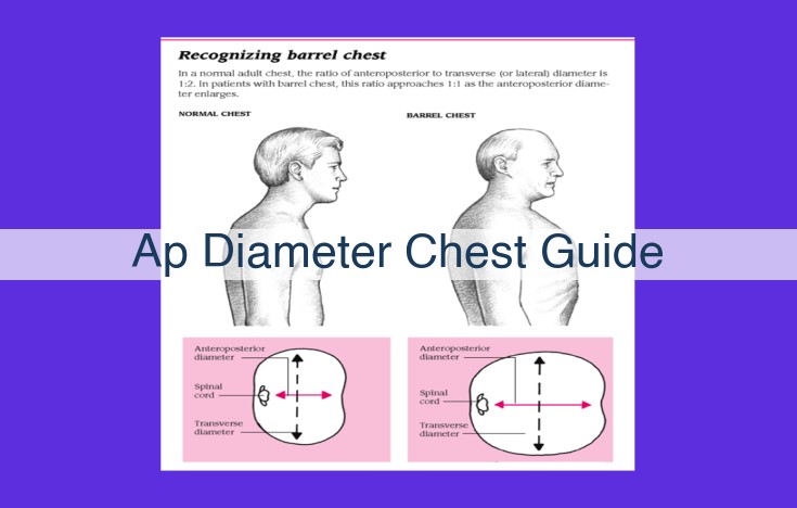
The AP diameter, measured in centimeters, represents the transverse dimension of the chest as seen on a chest radiograph. This measurement serves as a crucial indicator of the chest’s size and volume, providing insights into potential abnormalities.
Obtaining the AP diameter is an intricate process that demands precision and accuracy. It involves measuring the distance from the posterior aspect of the spine to the anterior chest wall, usually at the level of the vertebral bodies. This measurement is essential for evaluating the chest’s size, shape, and underlying structures.
Chest Radiograph: The Essential Tool for Measuring the AP Diameter
In the realm of radiology, the anteroposterior (AP) diameter plays a crucial role in assessing chest anatomy. The chest radiograph stands as an indispensable tool in obtaining this measurement, which provides vital insights into the health of the thoracic cavity.
The chest radiograph captures a detailed image of the chest, revealing the intricate structures within. Components of the chest radiograph include the mediastinum, lungs, heart, and bones. This comprehensive view allows radiologists to interpret the anatomy and identify any abnormalities.
Measuring the AP diameter on a chest radiograph is a precise process. By drawing a line from the posterior aspect of the vertebral body to the anterior chest wall, radiologists establish the AP diameter. This measurement serves as an indicator of the overall chest size and shape.
Deviations from the normal AP diameter can point to underlying medical conditions. For instance, an enlarged AP diameter may suggest pulmonary hyperinflation, while a decreased AP diameter could indicate a restrictive lung process.
The chest radiograph remains the gold standard for evaluating the AP diameter and detecting chest abnormalities. Its versatility and wide availability make it an essential tool in medical diagnosis and patient management.
AP Diameter: A Key Measurement for Chest Health
The anteroposterior (AP) diameter is a crucial measurement in assessing chest anatomy, especially on chest radiographs. It provides valuable insights into the overall size, shape, and symmetry of the chest cavity.
The AP diameter is measured on a chest radiograph (X-ray) taken in the posteroanterior (PA) position, where the X-ray beam passes through the body from back to front. The measurement is taken from the posterior aspect of the vertebral bodies to the anterior aspect of the sternum.
Importance in Detecting Chest Abnormalities
The AP diameter plays a significant role in detecting various chest abnormalities. For instance:
- An increased AP diameter may indicate conditions such as emphysema or pneumothorax, where the lungs become overinflated.
- A decreased AP diameter may suggest atelectasis, a condition where the lungs collapse, or kyphosis, an abnormal curvature of the spine.
- An asymmetric AP diameter may point to unilateral lung disease, such as fibrosis or pneumonia.
By evaluating the AP diameter in conjunction with other chest radiograph findings, healthcare professionals can identify and diagnose a wide range of chest conditions, including infections, masses, and structural abnormalities.
Standard PA Chest Film: Positioning and Alignment
Proper positioning and alignment are crucial for accurate measurement of the AP diameter.
The AP diameter chest guide provides guidelines for the correct positioning of the patient and alignment of the X-ray beam. The patient stands facing the X-ray tube with their chest against the cassette. The cassette should be centered over the spine.
The patient should be instructed to hold their breath at the end of expiration and keep their arms raised above their head. The X-ray beam is projected from the back, passing through the chest and exposing the cassette.
The AP diameter is measured from the posterior aspect of the vertebral bodies to the anterior aspect of the sternum. The measurement should be parallel to the costal margins. By following these guidelines, accurate and consistent measurements of the AP diameter can be obtained, aiding in the assessment of chest anatomy.
Anatomy of the Chest: Its Implications for AP Diameter
Understanding the anatomy of the chest is crucial for accurately interpreting the anteroposterior (AP) diameter, a measurement derived from chest radiographs that offers valuable insights into chest anatomy.
Cardiac Silhouette
The cardiac silhouette occupies a central position in the chest radiograph. Its location, morphology, and size provide crucial clues about the AP diameter. Normally, the cardiac silhouette should be symmetrical and well-defined. Any asymmetry or enlargement may indicate abnormalities, such as cardiomegaly, affecting the AP diameter.
Pleural Space
The pleural space is a potential space bounded by the visceral and parietal pleura. It serves as a cushion around the lungs and mediastinal structures. In chest radiographs, the pleural space appears as a thin, linear opacity on both sides of the cardiac silhouette. Widening of the pleural space can signify pleural effusion or pneumothorax, impacting the AP diameter measurements.
Mediastinum
The mediastinum is the central compartment of the chest, housing vital structures such as the heart, trachea, and great vessels. Its contents can influence the AP diameter. An enlarged mediastinum may compress the lungs, narrowing the AP diameter, while an anterior mediastinal mass may push the heart forward, increasing the AP diameter.
Vertebral Bodies
The vertebral bodies form the posterior boundary of the chest radiograph. They serve as anatomical landmarks for AP diameter measurements. The alignment and spacing of the vertebral bodies should be symmetrical. A shift or rotation of the vertebrae can alter the AP diameter measurements.
Pulmonary Artery
The pulmonary artery is a large vessel that carries blood from the heart to the lungs. Its size and location contribute to the cardiac silhouette’s appearance. Normally, the pulmonary artery should be relatively small compared to the aorta. Enlargement of the pulmonary artery may indicate pulmonary hypertension, potentially affecting the AP diameter.
Diaphragm
The diaphragm is the muscular sheet that separates the thoracic and abdominal cavities. Its elevation or descent can influence the AP diameter. A high diaphragm may reduce the AP diameter, while a low diaphragm may increase it. This elevation can result from conditions such as diaphragmatic hernia or phrenic nerve palsy.