Discover The Secrets Of Dental Anatomy: Cusps, Grooves, And The Art Of Mastication
The cusps of teeth are raised projections on the crown’s chewing surface. They may be buccal, facing the cheeks; lingual, facing the tongue; mesial, positioned towards the midline; or distal, located away from it. These surfaces are crucial for mastication, providing a gripping and grinding mechanism. Additionally, the incisal edge of anterior teeth (incisors and canines) performs a specialized cutting function. Grooves, ridges, pits, and fossae contribute to the unique morphology of teeth, aiding in food manipulation and retention. Understanding dental anatomy is essential for dentists in diagnosing and treating dental issues effectively, as well as for patient education purposes.
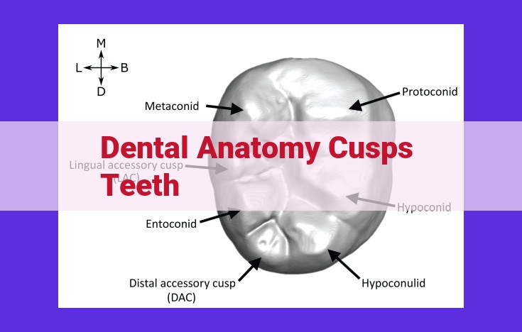
Types of Cusps: The Building Blocks of Teeth
Our teeth are intricate structures that play a vital role in our daily lives. Understanding their anatomy is essential for maintaining good oral health. Among the key anatomical features of teeth are cusps, the protruding points that give them their shape and function.
Four Primary Types of Cusps
Cusps come in four primary types: buccal, lingual, mesial, and distal. Each type has a specific location and function:
- Buccal Cusps: Located on the outside surface of the tooth, facing the cheek, buccal cusps help grind food.
- Lingual Cusps: Found on the inside surface of the tooth, facing the tongue, lingual cusps shear food.
- Mesial Cusps: Positioned on the front side of the tooth, toward the midline, mesial cusps grip and tear food.
- Distal Cusps: Situated on the back side of the tooth, away from the midline, distal cusps cut and pulverize food.
These cusps work in concert to create a rough, uneven surface that aids in the mastication process. Their interlocking pattern allows for efficient grinding, tearing, and cutting, ensuring that food is properly broken down for digestion.
Surfaces of Teeth: Navigating the Dental Landscape
Every tooth is a marvel of engineering, boasting a landscape of distinct surfaces that play crucial roles in our ability to eat, speak, and smile with confidence. Understanding the nuances of these surfaces is essential for dentists and other dental professionals to diagnose, treat, and educate their patients effectively.
1. Occlusal Surface: The Grinding Ground
The occlusal surface is the biting surface of a tooth that meets its counterpart in the opposing jaw. It’s designed to crush and grind food into digestible particles. Only the posterior teeth—molars and premolars—have this surface.
2. Facial Surface: A Front Profile
The facial surface is the front or cheek-facing surface of a tooth. It’s smooth and visible when we smile. All teeth, except the maxillary molars that have no facial surface, display this surface prominently.
3. Lingual Surface: The Tongue’s Domain
The lingual surface is the tongue-facing surface of a tooth. Just like the facial surface, it’s present in all teeth except the mandibular molars, which lack a lingual surface.
4. Mesial Surface: The Neighbor on the Left
The mesial surface is the surface that faces the midline of the mouth or the tooth next to it. It’s found on all teeth except the most posterior molars.
5. Distal Surface: The Neighbor on the Right
The distal surface is the surface that faces away from the midline of the mouth or the tooth next to it. Similar to the mesial surface, it’s present on all teeth except the most anterior incisors.
Understanding the Surfaces: A Key to Dental Health
Incisive knowledge of tooth surfaces empowers dentists with the ability to:
- Identify areas of decay and wear
- Plan fillings and other restorative procedures
- Effectively communicate oral hygiene techniques to patients
- Provide tailored advice on bite alignment and tooth grinding habits
By immersing ourselves in the dental landscape, we can appreciate the intricacies of our teeth and the fundamental role they play in our overall health and well-being.
The Incisal Edge: A Gateway to a Radiant Smile
When we think of pearly whites, our minds often conjure images of incisors, those front teeth that grace our smiles. These teeth boast a distinguishing feature—the incisal edge—a sharp, straight margin that forms the cutting surface of your mouth.
The incisal edge is designed for slicing and shearing, enabling us to effortlessly bite into food. Its blade-like precision allows us to tear through fibrous vegetables and slice fruits with ease. The central and lateral incisors, positioned at the center and sides of our dental arch, respectively, bear this incisal edge, making them the primary incisors responsible for our biting prowess.
Understanding the incisal edge goes beyond its function in mastication. For dentists, it serves as a crucial landmark in diagnosing and treating dental issues. By carefully examining the edge’s alignment, wear patterns, and any chips or fractures, dental professionals can assess the overall health of your teeth and accurately plan appropriate treatments.
So, next time you gaze at your reflection, take a closer look at your incisal edges. These unassuming yet essential features not only contribute to your radiant smile but also play a vital role in your dental well-being.
Grooves, Ridges, Fossae, and Pits
- Define each term and provide examples of where they can be found on teeth.
- Explain their role in the structure and function of teeth.
Grooves, Ridges, Fossae, and Pits: The Intricate Topography of Your Teeth
The surfaces of our teeth are not as smooth as they appear. A closer examination reveals a fascinating array of grooves, ridges, fossae, and pits that play crucial roles in the structure and function of our teeth.
Definition and Location:
- Grooves: Narrow, elongated depressions that run along the surfaces of teeth. They help guide the opposing teeth during chewing. Examples include the buccal groove on the cheek side of molars and the palatal groove on the lingual side of molars.
- Ridges: Elevated lines that run parallel to the grooves. They provide strength and support to the teeth, preventing excessive wear. Examples include the buccal ridge on the cheek side of molars and the palatal ridge on the lingual side of molars.
- Fossae: Broad, shallow depressions on the chewing surface of teeth. They accommodate the cusps of opposing teeth, providing a grinding area for food. Examples include the central fossa and distal fossa on the occlusal surface of molars.
- Pits: Small, deep depressions found at the intersections of grooves and ridges. They are often weak points where decay can start. Examples include the distolingual pit on the lingual side of molars and the mesiobuccal pit on the cheek side of molars.
Role in Structure and Function:
These anatomical features work in harmony to ensure the proper function of our teeth:
- The grooves and ridges provide a guide for chewing motions, preventing teeth from sliding off each other.
- The ridges strengthen the teeth, preventing excessive wear and damage.
- The fossae create a grinding area for food, helping to break it down into smaller pieces during chewing.
- The pits provide retention areas for food particles, making cleaning more challenging.
Understanding these anatomical features is crucial for maintaining good oral health. Dentists rely on this knowledge to:
- Diagnose dental problems such as cavities and fractures.
- Plan and perform dental treatments such as fillings and crowns.
- Educate patients about the importance of proper oral hygiene.
By appreciating the intricate topography of our teeth, we can better understand their function and take better care of our smiles.
Clinical Significance of Dental Anatomy
Diagnosis and Treatment:
For dental professionals, a solid understanding of dental anatomy serves as a cornerstone for accurate diagnosis and effective treatment. Identifying the specific surfaces, cusps, and grooves allows dentists to pinpoint the location of caries, trauma, or other dental issues with precision. This detailed knowledge guides decision-making for restorative procedures, such as fillings, crowns, and root canals, ensuring optimal outcomes for patients.
Patient Education:
Dental anatomy also plays a crucial role in patient education. When dentists explain the structure and function of teeth, it empowers patients to better understand their own oral health. This knowledge fosters a sense of responsibility and encourages preventive measures, such as proper brushing and flossing techniques, reducing the risk of future dental problems.
Communication and Collaboration:
Clear communication is essential within the dental team. A shared understanding of dental anatomy facilitates seamless collaboration between dentists, hygienists, and other professionals. Accurate terminology and standardized descriptions allow for precise communication regarding treatment plans, referrals, and dental records, ensuring consistent and high-quality patient care.
Continuing Education:
Dental anatomy is a foundation upon which all other aspects of dentistry rest. Ongoing education and advancements in the field require a solid grasp of this foundational knowledge. Attending conferences, workshops, and reading scientific literature allows dental professionals to stay abreast of the latest techniques and technologies, maximizing their ability to provide exceptional patient care.