Emg Monitoring In Muscle Relaxant Optimization: Insights For Anesthesiologists And Clinicians
EMG results provide valuable insights into the effectiveness of muscle relaxers. EMG monitors reduced muscle activity, decreased muscle tone, lower EMG amplitude, fewer muscle contractions, and prolonged muscle relaxation. The key EMG parameters include reduced EMG amplitude, increased inter-spike intervals, and decreased spontaneous activity. By interpreting these results, anesthesiologists and clinicians can optimize muscle relaxant administration during anesthesia and medical procedures, ensuring patient safety and optimal outcomes.
Decoding the Language of Muscles: How EMG Illuminates the Effects of Muscle Relaxers
Imagine a concert where the performers are your muscles. Electromyography (EMG) acts as the conductor, capturing the electrical signals that orchestrate every muscle movement. When you introduce muscle relaxants into this symphony, EMG becomes an invaluable tool for understanding their impact on your muscular ensemble.
Muscle Relaxers: A Journey into Muscular Slumber
Muscle relaxers are like gentle whispers that lull your muscles into a state of tranquility. They work by blocking the communication pathways between nerves and muscles, effectively putting your muscles into a temporary slumber. This slumberous state is crucial for surgeries and other medical procedures, allowing surgeons to work with minimal muscle interference.
EMG: The Window into Muscle Dialogue
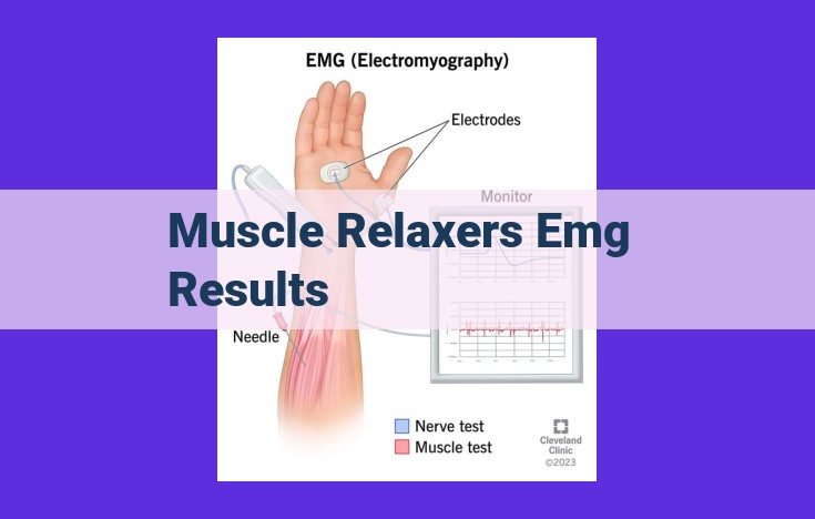
EMG is the keyhole through which we peer into the muscular world. It records the electrical chatter of your muscles, revealing their activity levels, tone, and the timing of their contractions. By analyzing these electrical conversations, we can gauge the potency of muscle relaxers and ensure their safe and effective use.
Reduced Muscle Activity: How Muscle Relaxers Inhibit Muscle Function
In the realm of anesthesia and medical procedures, muscle relaxers play a crucial role in immobilizing the patient for optimal conditions. These medications essentially dampen the body’s muscle activity, making it easier to perform surgical interventions without involuntary movements. To effectively evaluate the effectiveness of these muscle relaxants, electromyography (EMG) emerges as a valuable tool. EMG monitors electrical activity in muscles, providing insights into their behavior under the influence of these medications.
When muscle relaxants are administered, EMG reveals telltale signs of reduced muscle activity. These key parameters provide an objective measure of the drug’s impact on muscle function.
-
Decreased EMG Amplitude: EMG measures the strength of electrical signals generated by muscles. Under the effect of muscle relaxants, EMG amplitude diminishes, reflecting a reduction in muscle activity. This diminished electrical activity corresponds with the drug’s ability to interfere with nerve impulses that trigger muscle contractions.
-
Fewer Muscle Contractions: EMG also detects the frequency of muscle contractions. As muscle relaxants take effect, the number of muscle contractions decreases. This observation aligns with the drug’s mechanism of action, blocking the signals that initiate muscle movements. The diminished contractions indicate the successful inhibition of muscle activity, ensuring optimal conditions for medical procedures.
-
Prolonged Inter-Spike Intervals: EMG records the time between successive electrical spikes, known as inter-spike intervals. When muscle relaxants are present, inter-spike intervals lengthen, indicating a slowdown in muscle activity. This parameter offers additional evidence of the drug’s ability to impede nerve impulses responsible for muscle contractions.
By monitoring these EMG parameters, clinicians can accurately assess the level of muscle relaxation induced by the drug. This information guides clinical decision-making, ensuring adequate muscle relaxation for the procedure while minimizing the risk of excessive paralysis.
Decreased Muscle Tone: Unveiling the Impact of Muscle Relaxers through EMG
When it comes to understanding the effects of muscle relaxers, electromyography (EMG) plays a crucial role. By analyzing electrical signals from muscles, EMG provides valuable insights into how these medications alter muscle activity and tone.
Muscle Tone and EMG Amplitude
Muscle tone refers to the constant, low-level muscle contraction that keeps our bodies upright and poised. EMG amplitude, which measures the electrical activity of muscles, is directly related to muscle tone. Higher EMG amplitude indicates greater muscle activity and tone.
Impact of Muscle Relaxers on Muscle Tone
Muscle relaxers, as their name suggests, reduce muscle tone. They work by blocking the transmission of nerve impulses to muscles, thereby inhibiting muscle contractions. This results in a decrease in EMG amplitude, reflecting the diminished muscle activity and tone.
Implications for Clinical Practice
Monitoring EMG amplitude during muscle relaxant administration is essential in clinical practice. By tracking the changes in muscle tone, healthcare providers can optimize dosage and ensure safe and effective muscle relaxation. For instance, in anesthesia, EMG helps guide the administration of muscle relaxants to achieve the desired level of muscle paralysis for intubation and surgical procedures.
EMG provides a valuable window into the impact of muscle relaxers on muscle tone. By analyzing EMG amplitude, healthcare professionals can accurately assess the effectiveness of muscle relaxation and make informed decisions regarding medication administration. This ensures optimal outcomes for patients undergoing procedures that require muscle relaxation.
Lower EMG Amplitude: Unveiling Muscle Relaxant Effects
EMG: A Window into Muscle Activity
EMG (electromyography) is a diagnostic tool that measures electrical activity in muscles. It provides valuable insights into muscle function and the effects of medications like muscle relaxants.
Reduced EMG Amplitude: A Tale of Inhibited Muscle Contractions
When muscle relaxants take effect, they inhibit neurotransmission at the neuromuscular junction, the point where nerves communicate with muscles. This interference hinders the generation and propagation of electrical signals within muscles.
As a result, EMG recordings reveal a reduced amplitude of muscle activation. The lower the EMG amplitude, the less active the muscle becomes.
Correlation with Muscle Strength
EMG amplitude directly correlates with muscle strength. A lower EMG amplitude indicates weaker muscle contractions. This is because reduced electrical activity translates into fewer and weaker muscle fibers being activated.
Clinical Implications: Monitoring Muscle Weakness
EMG monitoring during anesthesia and surgical procedures plays a crucial role in assessing the efficacy and safety of muscle relaxants. By tracking EMG amplitude, clinicians can optimize dosing to ensure adequate muscle relaxation without causing excessive weakness.
Fewer Muscle Contractions: Unveiling Clinical Implications
EMG’s Astute Detection of Decreased Muscle Contractions
Electromyography (EMG) plays a pivotal role in detecting the subtle decline in muscle contractions brought on by muscle relaxants. Its ability to capture electrical impulses emitted by muscles allows clinicians to monitor muscle activity with precision. As muscle relaxants take hold, EMG records a noticeable reduction in these electrical signals, indicating a corresponding decrease in muscle contractions.
Clinical Significance of Reduced Muscle Contractions
The clinical implications of reduced muscle contractions extend beyond the operating room. In anesthesia, precise monitoring ensures adequate muscle relaxation for safe intubation and surgical procedures. It helps prevent coughing, laryngospasm, and other complications that can arise from incomplete muscle relaxation. Conversely, over-relaxation can lead to respiratory depression, highlighting the crucial role of EMG in optimizing muscle relaxant administration.
EMG as a Guide for Clinical Decision-Making
By interpreting EMG signals, clinicians can determine the appropriate dosage and timing of muscle relaxants. EMG provides real-time feedback, allowing them to adjust medication administration to achieve the desired level of muscle relaxation. This meticulous monitoring helps prevent complications associated with both under- and over-relaxation, ensuring patient safety.
EMG in Other Medical Scenarios
EMG’s utility extends beyond anesthesia. It assists in diagnosing and managing conditions like myasthenia gravis, where muscle weakness results from reduced acetylcholine release. EMG can also monitor muscle function during neuromuscular junction disorders, aiding in proper diagnosis and treatment.
EMG plays a vital role in monitoring muscle relaxant effects, ensuring safe and effective use in medical procedures. Its ability to detect fewer muscle contractions provides valuable clinical insights, guiding optimal dosage and timing of medication administration. EMG empowers clinicians to deliver personalized care, reducing risks and improving patient outcomes.
Prolonged Muscle Relaxation: Unraveling the Secrets of Muscle Relaxers
Identifying Prolonged Muscle Relaxation with EMG
Electromyography (EMG) plays a crucial role in monitoring the effects of muscle relaxants during anesthesia and medical procedures. When muscle relaxers effectively inhibit muscle activity, EMG parameters reveal specific patterns that indicate prolonged muscle relaxation.
EMG measures the electrical activity of muscles. During prolonged muscle relaxation, the following EMG parameters provide valuable insights:
-
Reduced EMG Amplitude: A decrease in the amplitude (height) of the EMG waveform indicates a significant reduction in muscle activity. This finding suggests that the muscle relaxant has successfully blocked the transmission of nerve impulses to the muscles.
-
Lower EMG Frequency: The frequency of EMG signals refers to the number of muscle contractions per second. Prolonged muscle relaxation leads to a lower EMG frequency, indicating a decreased number of muscle contractions.
-
Increased Inter-Spike Intervals: EMG waveforms exhibit spikes that represent individual muscle fiber contractions. During prolonged muscle relaxation, the inter-spike intervals (the time between spikes) increase. This indicates a slower rate of muscle fiber firing and further inhibition of muscle activity.
Mechanism of Action: How Muscle Relaxers Promote Prolonged Muscle Relaxation
Muscle relaxants achieve prolonged muscle relaxation by interfering with the neuromuscular junction, the site where nerve impulses trigger muscle contractions. There are two main types of muscle relaxants:
-
Non-depolarizing Muscle Relaxants: These agents block the acetylcholine receptors on muscle cells, preventing nerve impulses from causing muscle contraction.
-
Depolarizing Muscle Relaxants: These agents act by continuously depolarizing the muscle cell membrane, making it resistant to nerve impulses and blocking muscle contraction.
Both types of muscle relaxants can produce prolonged muscle relaxation when administered in appropriate doses. The duration of muscle relaxation depends on the potency of the relaxant, the patient’s individual response, and the presence of any underlying conditions that may affect muscle function.
Increased Inter-Spike Intervals: Unveiling the Impact of Muscle Relaxers on Nerve Communication
In the realm of muscle relaxation, EMG (Electromyography) emerges as an invaluable tool, providing a window into the intricate workings of muscles. Among the myriad parameters measured by EMG, inter-spike intervals hold particular significance in deciphering the effects of muscle relaxants.
Inter-spike intervals, measured in milliseconds, represent the time elapsed between successive electrical impulses generated by muscle fibers. Under normal conditions, these intervals are relatively consistent, reflecting the rhythmic contractions of the muscle. However, when muscle relaxants enter the scene, their molecular dance with nerve signals disrupts this delicate rhythm, resulting in prolonged inter-spike intervals.
This elongation of inter-spike intervals is a telltale sign of reduced nerve-muscle communication. Muscle relaxants exert their influence by binding to receptors on the surface of muscle fibers, effectively blocking the transmission of nerve impulses. With fewer signals reaching their targets, muscle activity dwindles, and the inter-spike intervals stretch out.
The Significance of Prolonged Inter-Spike Intervals
Prolonged inter-spike intervals are not merely academic curiosities; they have profound clinical implications. By monitoring these intervals, anesthesiologists and healthcare professionals can gauge the depth of muscle relaxation induced by the administered relaxant.
In the operating room, maintaining adequate muscle relaxation is paramount for ensuring patient safety and facilitating surgical procedures. Over-relaxation can lead to respiratory depression, while under-relaxation may hinder surgical visibility and increase the risk of patient movement. EMG monitoring, through its detection of prolonged inter-spike intervals, provides real-time feedback on the effects of muscle relaxants, allowing clinicians to tailor their administration accordingly.
Unveiling the Molecular Mechanisms
The mechanisms by which muscle relaxants prolong inter-spike intervals are fascinating. These drugs primarily target acetylcholine, a neurotransmitter that plays a pivotal role in muscle contraction. By competing with acetylcholine for binding sites on muscle fibers, muscle relaxants effectively block the transmission of nerve impulses.
This blockade disrupts the normal depolarization-repolarization cycle of muscle fibers, resulting in reduced muscle activity and the prolongation of inter-spike intervals. The extent of interval prolongation depends on the type and dosage of muscle relaxant used, as well as the individual patient’s sensitivity to the drug.
In the symphony of muscle relaxation, inter-spike intervals serve as a conductor, orchestrating the balance between adequate relaxation and patient safety. By understanding the impact of muscle relaxers on these intervals, healthcare professionals can harness the power of EMG monitoring to optimize muscle relaxant administration, ensuring a smooth and uneventful course of anesthesia and medical procedures.
Interpretation of EMG Results
When interpreting EMG results for muscle relaxants, healthcare professionals rely on key parameters that reflect the drug’s effects. These parameters include:
- Reduced Muscle Activity: EMG shows a decrease in muscle activity, indicated by fewer and smaller action potentials.
- Decreased Muscle Tone: EMG amplitude, which measures the electrical activity of muscles, decreases due to reduced muscle tone.
- Lower EMG Amplitude: The average voltage of EMG signals diminishes, signifying less muscle activity.
- Fewer Muscle Contractions: EMG detects fewer muscle contractions, indicating decreased muscle function.
- Prolonged Muscle Relaxation: EMG shows persistent muscle relaxation, evidenced by prolonged periods of electrical silence.
- Increased Inter-Spike Intervals: The time between successive action potentials increases, indicating impaired muscle transmission.
These EMG parameters guide clinical decision-making by providing insights into the depth and duration of muscle relaxation. Healthcare professionals can optimize muscle relaxant administration based on these findings, ensuring adequate relaxation for necessary medical procedures while minimizing the risk of complications.
Clinical Applications of EMG in Muscle Relaxant Management
EMG, paired with muscle relaxants, plays a pivotal role in the safe and effective conduction of numerous medical procedures. In anesthesia, EMG aids in monitoring muscle relaxation, ensuring adequate muscular blockade for surgical interventions. It enables anesthesiologists to precisely titrate muscle relaxant administration, optimizing surgical conditions while minimizing potential complications.
Furthermore, EMG is crucial in other medical settings where muscle relaxation is necessary, such as endoscopy, radiology, and orthopedic procedures. By providing real-time feedback on muscle activity, EMG ensures appropriate muscle relaxation for these delicate procedures.
In the clinical arena, EMG results guide informed decision-making regarding muscle relaxant administration. For instance, analyzing EMG parameters helps clinicians assess the depth of muscle relaxation. They can then adjust muscle relaxant dosages to maintain optimal surgical conditions without overdosing, which could lead to adverse effects such as prolonged paralysis.
Moreover, EMG plays a vital role in extubation. By evaluating EMG activity, clinicians can determine the readiness of patients to breathe spontaneously after surgery. This reduces the risk of postoperative complications, such as pulmonary aspiration.