Dna Sizing Standards: Challenges, Innovations, And Multifaceted Approaches
Unveiling the mysteries surrounding DNA size standards, this article delves into the challenges and limitations of traditional methods, exploring PCR amplification bias, GC bias, and innovative technologies like optical mapping and nanopore sequencing. It highlights the need for multifaceted approaches to ensure accurate DNA fragment analysis, combining multiple techniques and computational tools. Ongoing research and advancements in this field promise to further refine our understanding and pave the way for more precise and reliable DNA size determinations.
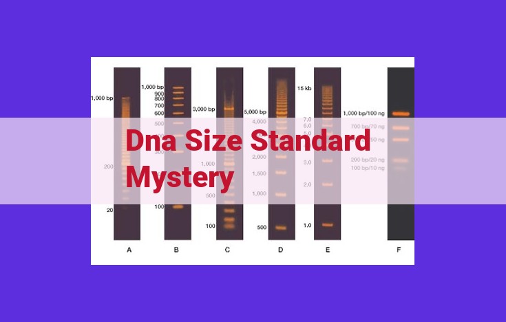
DNA Size Standards: The Cornerstone of Fragment Analysis
In the intricate world of DNA analysis, a critical foundation stone lies in the humble DNA size standards. These remarkable tools serve as the measuring sticks for determining the length of DNA fragments, enabling researchers to unravel the secrets of genetic material.
DNA size standards are meticulously designed DNA fragments of precisely known lengths. By comparing the migration patterns of unknown DNA fragments to these standards, scientists can accurately estimate the sizes of the unknown fragments.
There exists a diverse array of DNA size standards, each tailored to specific applications. Molecular weight markers, for instance, provide broad coverage across a wide range of fragment sizes. Single-stranded DNA size standards, on the other hand, excel in resolving small fragments.
Despite their invaluable role, traditional DNA size standards possess inherent challenges. Variations in fragment composition and sequence can introduce biases, leading to inaccuracies in size estimation. Additionally, traditional methods may struggle to resolve fragments of similar sizes or fragments that exhibit unusual melting behavior.
PCR Amplification Bias: A Hidden Obstacle
- Explain the principles of polymerase chain reaction (PCR) and its role in DNA analysis.
- Explore the phenomenon of PCR amplification bias and its impact on DNA fragment size estimation.
- Provide strategies to mitigate the effects of PCR amplification bias.
PCR Amplification Bias: Unmasking a Hidden Obstacle in DNA Analysis
Polymerase chain reaction (PCR) is a revolutionary technique widely employed in DNA analysis. It allows scientists to amplify specific DNA sequences, creating millions of copies for further analysis. However, a hidden obstacle lurking within PCR is amplification bias. This phenomenon can significantly impact DNA fragment size estimation, leading to inaccurate results.
Amplification bias arises from the unequal amplification of different DNA fragments during PCR. Some sequences amplify more efficiently than others, depending on their length, GC content, and secondary structure. As a result, the relative abundance of different fragment sizes in the amplified product may not accurately reflect their true proportions in the original sample.
This bias can have serious consequences in DNA size analysis, especially when using techniques that rely on the accurate estimation of fragment sizes, such as capillary electrophoresis or next-generation sequencing. Incorrect size estimation can lead to misinterpretation of data and flawed conclusions.
To mitigate the effects of PCR amplification bias, researchers have developed several strategies:
- Optimizing PCR conditions: By adjusting factors such as primer design, annealing temperature, and polymerase choice, scientists can improve the unbiased amplification of different DNA fragments.
- Using internal size standards: Internal size standards, which are DNA fragments of known size, can be added to the PCR reaction to calibrate the size estimation during data analysis.
- Normalizing PCR products: Quantifying the amount of amplified DNA and then normalizing the input for size analysis can help reduce the impact of amplification bias.
Understanding and overcoming PCR amplification bias is crucial for accurate DNA fragment size estimation. By employing appropriate strategies, researchers can ensure the integrity of their data and enhance the reliability of their DNA analysis.
Understanding GC Bias: The Influence of Nucleotide Composition on DNA Fragment Size
In the realm of DNA analysis, the accuracy of determining fragment sizes is crucial. However, a hidden obstacle lurking in this process is GC bias, a phenomenon influenced by the nucleotide composition of DNA fragments.
GC bias arises from the varying melting temperatures of DNA regions with different ratios of guanine (G) and cytosine (C) nucleotides. Fragments with higher GC content melt at higher temperatures due to the formation of stronger hydrogen bonds between G-C base pairs. This thermal instability affects the efficiency of both polymerase chain reaction (PCR) amplification and enzymatic fragmentation.
During PCR, DNA polymerases may favor amplifying DNA regions with lower GC content, resulting in unequal amplification of fragments with different GC compositions. This bias can lead to overestimation of fragment sizes.
Enzymatic fragmentation, commonly used to prepare DNA for analysis, can also be influenced by GC bias. Restriction enzymes, which cut DNA at specific recognition sequences, may exhibit preferential cleavage at AT-rich regions, leading to the underestimation of fragment sizes.
Addressing GC bias is essential to ensure accurate fragment size estimation. Techniques like PCR optimization and the use of high-fidelity polymerases can help minimize amplification bias. Enzymatic fragmentation optimization and the employment of alternative restriction enzymes with reduced GC sensitivity can also mitigate size underestimation.
Researchers are actively exploring novel approaches to overcome GC bias. Computational tools are being developed to correct for GC-induced distortions. Future advancements in sequencing technologies, such as long-read sequencing, hold promise for direct and unbiased size determination.
By understanding and addressing GC bias, scientists can enhance the accuracy and precision of DNA fragment size analysis, paving the way for reliable and informative genetic studies.
Optical Mapping: Unveiling Fragment Sizes Directly
- Describe the principles and methodology of optical mapping for DNA fragment analysis.
- Highlight the advantages and disadvantages of optical mapping compared to traditional size standards.
- Compare and contrast the accuracy and efficiency of optical mapping.
Optical Mapping: Unveiling Fragment Sizes Directly
Optical mapping, a revolutionary technique in DNA analysis, offers a unique perspective on DNA fragment sizing, paving the way for unprecedented accuracy and efficiency. Unlike traditional methods that rely on size standards, optical mapping employs a direct approach, capturing the physical contours of DNA molecules.
Principles and Methodology
Optical mapping harnesses a high-resolution microscope to scan and image individual DNA molecules. The DNA is first stretched and stained with fluorescent dyes, allowing the microscope to visualize and capture its intricate structure. Specialized software then analyzes the resulting images, identifying and measuring DNA fragments based on their unique patterns.
Advantages over Traditional Size Standards
Compared to traditional size standards, optical mapping boasts several advantages. It provides:
- Higher Accuracy: By directly measuring DNA molecules, optical mapping eliminates the inherent inaccuracies associated with size standards.
- Wide Range of Fragment Sizes: Optical mapping can accurately size fragments ranging from hundreds of base pairs to megabases, covering a much wider range than traditional methods.
- Complex Structure Analysis: Optical mapping also unveils the structural complexities of DNA, such as gaps, deletions, and inversions, which can be missed by standard techniques.
Disadvantages
However, optical mapping is not without its limitations:
- Cost and Expertise: The equipment and expertise required for optical mapping can be costly and specialized.
- Time-Consuming: The process of imaging and analyzing DNA molecules can be time-consuming compared to standard methods.
- Sample Preparation: DNA samples must be carefully prepared to ensure optimal visualization and accuracy.
Accuracy and Efficiency
In terms of accuracy, optical mapping surpasses traditional size standards. It provides highly precise measurements, even for complex and large DNA fragments. While not as fast as standard techniques, optical mapping offers significantly improved accuracy and a broader range of applications.
Optical mapping is a game-changer in DNA fragment analysis, offering a direct and accurate method to unveil the molecular details of DNA. By overcoming the limitations of traditional size standards, optical mapping opens up new possibilities in genetics, genomics, and other advanced biological research. As technology continues to advance, optical mapping promises to play an increasingly pivotal role in unlocking the secrets of the human genome and beyond.
Nanopore Sequencing: A Revolutionary Approach
- Introduce nanopore sequencing technology and its unique capabilities in DNA analysis.
- Explain how nanopore sequencing can be utilized for DNA size determination.
- Discuss the advantages and limitations of nanopore sequencing in this context.
Nanopore Sequencing: A Revolutionary Approach to DNA Size Determination
Advancements in DNA analysis methods have revolutionized our understanding of genetic information. In recent years, nanopore sequencing has emerged as a groundbreaking technology that is transforming the way we measure DNA fragment sizes. Here’s how this cutting-edge technique is revolutionizing DNA size determination:
Understanding Nanopore Sequencing
Nanopore sequencing harnesses the power of nanopores, tiny biological channels that allow ions to pass through them. By threading DNA molecules through these nanopores, researchers can detect the changes in electrical current caused by each nucleotide, the building block of DNA. This allows them to determine the sequence of the DNA fragment and its length.
Applications in DNA Size Determination
Nanopore sequencing offers a unique advantage in DNA size determination. Unlike traditional methods that rely on size standards or gel electrophoresis, nanopore sequencing directly measures the length of DNA fragments without the need for calibration or estimation. This eliminates potential errors and uncertainties associated with these traditional methods.
Advantages of Nanopore Sequencing
Nanopore sequencing offers several advantages over traditional size determination techniques:
- High accuracy: Direct measurement of DNA length ensures precise size determination.
- Long-read capability: Nanopore sequencing can analyze long DNA fragments (over 100,000 base pairs), overcoming the limitations of short-read sequencing technologies.
- Versatile applications: Nanopore sequencing is applicable to a wide range of DNA analysis scenarios, including genome assembly, structural variation detection, and DNA methylation mapping.
Limitations of Nanopore Sequencing
While nanopore sequencing is a powerful tool, it has certain limitations:
- Higher error rate: Nanopore sequencing technology is still prone to higher error rates compared to short-read sequencing methods.
- Long analysis time: Nanopore sequencing can take longer to generate results compared to some short-read sequencing platforms.
Nanopore sequencing is a revolutionary technology that is rapidly transforming the field of DNA size determination. Its ability to accurately and efficiently measure DNA fragment lengths has made it an invaluable tool for researchers in genetics, genomics, and precision medicine. As the technology continues to evolve and improve, nanopore sequencing promises to further revolutionize our understanding of genetic information.
Unveiling the Truth: A Multifaceted Approach to DNA Size Standard Analysis
The quest for accurate DNA size determination is a multifaceted endeavor, necessitating a combination of techniques and a keen understanding of the challenges that arise from traditional methods. DNA size standards serve as the foundation for fragment analysis, providing a reference point against which fragment sizes can be compared. However, these standards face inherent limitations, including potential bias introduced during DNA amplification and enzymatic fragmentation.
PCR amplification bias, a common obstacle, can arise from the preferential amplification of certain DNA fragments over others, leading to an overestimation of their size. GC bias, another factor to consider, refers to the influence of the nucleotide composition of a DNA fragment on its size. DNA fragments with a higher GC content tend to appear larger than fragments with a lower GC content.
To mitigate these challenges, researchers have turned to innovative technologies such as optical mapping and nanopore sequencing. Optical mapping offers a direct method for determining fragment sizes by creating a high-resolution map of the DNA molecule. Nanopore sequencing is a revolutionary approach that allows for the interrogation of DNA fragments in real-time, providing both size information and sequence data.
The combination of multiple techniques is crucial to unraveling the complexities of DNA size standard analysis. Computational tools play a vital role in data analysis and interpretation, ensuring accuracy and eliminating potential biases. Unveiling the truth requires an understanding of the limitations of each technique and the judicious integration of complementary approaches.
As research continues, future directions and ongoing challenges will shape the validation of DNA size standards. The development of new technologies and the refinement of existing methods hold promise for enhancing accuracy and efficiency. The quest for reliable DNA size determination is an ongoing endeavor, driven by the pursuit of truth and the advancement of scientific knowledge.